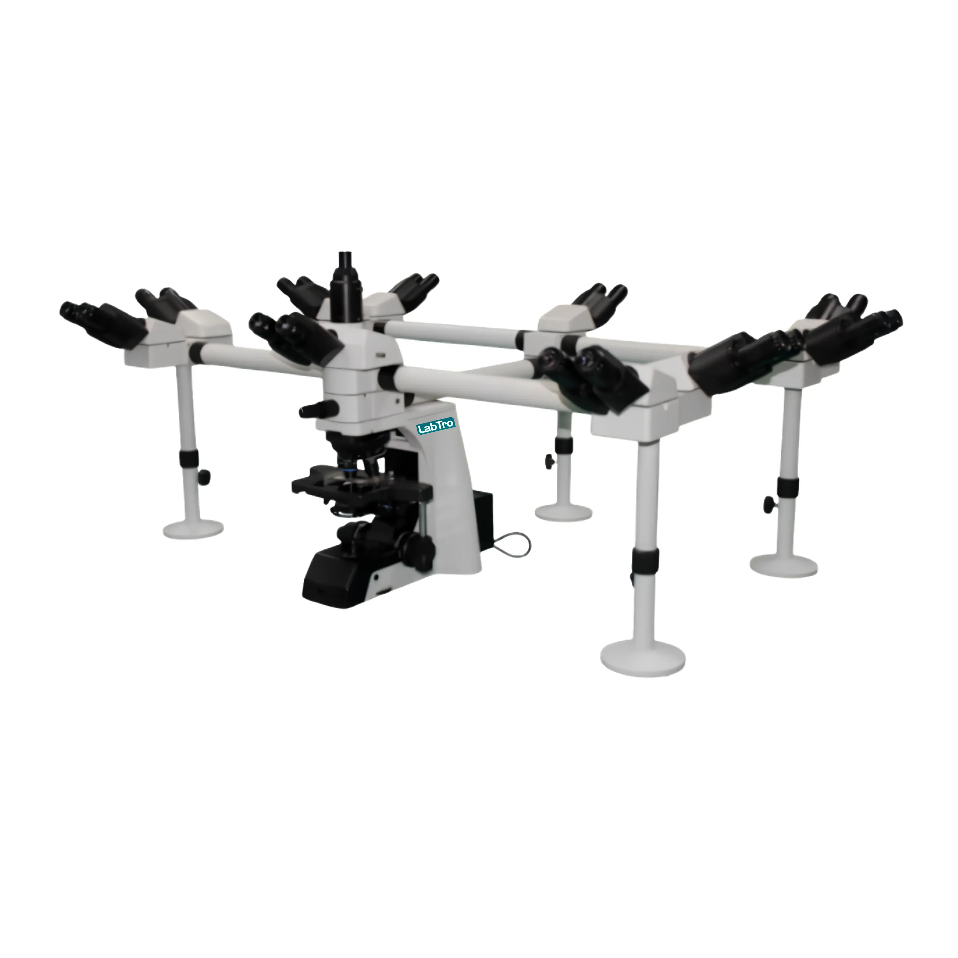

Multi-Viewing Microscope LT-MVM102

Multi-Viewing Microscope LT-MVM102

Labtro Multi-Viewing Microscope LT-MVM102 enables multiple users to observe the same specimen simultaneously through one trinocular and multiple binocular viewing heads. Its NIS60 infinite color-corrected optical system directs the magnified image through N-PLN Plan objectives from 4× to 100×, while the swing-out condenser controls illumination across the specimen. Low-position coaxial coarse and fine focusing with 0.001 mm precision allows accurate image adjustment, and the double-layer mechanical stage supports controlled slide movement. A 3 W adjustable S-LED illumination system provides consistent transmitted light during observation. The dual-color red and green LED pointer helps indicate specific specimen areas to all viewers, while the trinocular head supports connection to compatible digital imaging accessories for documentation and analysis.

Qty:

$ 11,000.00

Specifications

Optical System NIS60 Infinite Color Corrected Optical System
Main Viewing Head 1 × Seidentopf Trinocular Head
Main Head Inclination 30°
Main Head Interpupillary Distance 47 to 78 mm
Optical Splitting Ratio Eyepiece : Trinocular = 100:0 / 20:80 / 0:100
Additional Viewing Heads 9 × Seidentopf Binocular Heads
Binocular Head Inclination 30°
Binocular Head Interpupillary Distance 47 to 78 mm
Eyepieces 20 × Super Wide Field Plan Eyepieces SW10×/25 mm
Diopter Adjustment Adjustable
Nosepiece Backward Sextuple Nosepiece
DIC Slot Provided
Standard Objectives N-PLN Plan Objectives 4×, 10×, 20×, 40×, 100×
Optional Phase Contrast Objectives N-PLN PH 10×, 20×, 40×, 100×
Optional Fluorescence Objectives N-PLFN Plan Semi-Apochromatic 4×, 10×, 20×, 40×, 100×
Condenser Swing-Out Type Condenser
Condenser NA 0.9 / 0.25
Focusing System Low-position coaxial coarse and fine focusing
Fine Focus Division 1 µm / 0.001 mm
Focusing Moving Range 35 mm
Stage Type Double-layer Mechanical Stage
Stage Size 190 × 152 mm
Stage Moving Range 78 × 54 mm
Slide Holder Double slide holder
Stage Handle Right or left handle
Stage Precision 0.1 mm
Transmitted Illumination 3 W S-LED
Illumination Control Center pre-set, intensity adjustable
Optional Illumination 12 V / 100 W Halogen Lamp
Pointer Dual-color LED Pointer
Pointer Colors Green and Red
Pointer Brightness Adjustable
Photo Adapter Optional, for Nikon or Canon DSLR camera
Optional C-Mount Adapter 0.5× C-Mount, 1× C-Mount
Optional DIC Objectives 10× and 20× DIC Objective Lenses
Optional DIC Components Polarizer, DIC Insert Plates, DIC Turret Condenser
Optional Fluorescence Illuminator 6-position filter cube turret
Optional Fluorescence Filters B, G, U, V, R
Optional Mercury Lamp 100 W
Optional Fluorescence Power Controller 100 to 240 VAC
Optional Filters ND6 / ND25
Dust Cover Standard
Power Cord Standard
Cedar Oil 5 mL, Standard
Optional Polarizing Kit Simple Polarizing Kit
Optional Calibration Slide 0.01 mm
Optional Multi-Viewing Attachment 2 / 3 / 5 / 7 / 10 persons
Package size 77X54X51 cm
Gross weight 25 kg

Features

  • Multi-viewing optical system
    ↳ Allows multiple users to observe the same specimen simultaneously
  • NIS60 infinite color-corrected optics
    ↳ Supports clear and consistent image formation across the viewing heads
  • Plan objective configuration
    ↳ Includes N-PLN 4×, 10×, 20×, 40× and 100× objectives
  • Super-wide field eyepieces
    ↳ SW10×/25 mm eyepieces provide a broad viewing field with diopter adjustment
  • Precision focusing system
    ↳ Low-position coaxial coarse and fine focusing with 0.001 mm fine division
  • Mechanical stage control
    ↳ Double-layer stage with 78 × 54 mm movement supports accurate specimen positioning
  • Adjustable S-LED illumination
    ↳ 3 W transmitted illumination provides controlled brightness during observation
  • Dual-color LED pointer
    ↳ Red and green pointer helps indicate specific specimen areas to all viewers
  • Trinocular imaging capability
    ↳ Supports connection to compatible camera and digital imaging accessories
  • Expandable optical configuration
    ↳ Optional DIC, fluorescence, phase-contrast, polarizing and C-mount accessories extend functionality

Application

  • Pathology Laboratories → Group examination and discussion of tissue specimens
  • Cytology Laboratories → Collaborative observation of cellular morphology
  • Histology Laboratories → Review of prepared tissue sections by multiple users
  • Clinical Laboratories → Joint analysis of biological and diagnostic specimens
  • Medical Colleges → Instructor-led microscopy teaching and demonstrations
  • Research Institutes → Multi-user evaluation and comparison of research samples
  • Training Laboratories → Guided specimen observation using the dual-color pointer system

Expert Help Desk